


The human brain consists of billions of neurons connected to each other. To understand their work, scientists create a map of such connections – a connectome. We will tell you why it is important to study it and what projects exist in this area
Connectome and connectomics: what is it
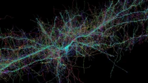
The brain is a complex organ. It consists of neurons that are connected to each other. At the points of their contact ( synapses ) there are small gaps. The synapses themselves are structures that can be seen with powerful electron microscopes.
When one neuron wants to “pass a message” to another, it releases special chemicals called neurotransmitters. They cross the synaptic gap and affect the neighboring cell, causing an electrical signal in it. This is how neurons exchange information: in this way, they influence thinking, emotions, and behavior.
For centuries, researchers have studied the structure of the brain, its cellular composition and biochemical properties. But the organization of connections between different areas remained a mystery until recently. The development of computer technology, artificial intelligence (AI) and big data analysis methods made it possible to solve this problem. A new field of knowledge emerged: connectomics.
The science of brain mapping (the process of creating detailed maps of brain regions) addresses how individual neurons and their associations interact with each other to influence thoughts and feelings.
The main focus of this science is the connectome . This is a map of neural pathways and connections in the brain. The connectome can be compared to the wiring diagram in a technical device, only much more complicated.
The term was proposed in 2005 by scientists Olaf Sporns, Rolf Ketter, Giulio Tononi and Patrick Hagmann. The concept was formed by analogy with the genome. If the genome is a complete set of genes, then the connectome is a complete map of neural connections. Scientists have found that it is unique for each person. The connectome differs even in two genetically identical people – identical twins.
A detailed map of all the connections in the human brain has not yet been compiled. This is due to the fact that this organ is very complex. It consists of approximately 86 billion neurons that form 100 trillion connections with each other. To map them, it is necessary to analyze a huge amount of data. Currently, scientists study the brain by compiling connectomes of individual areas of the cerebral cortex.
Neuroscientist Sebastian Seung, a professor at Princeton University’s Institute for Neuroscience, also noted that the map of connections changes over time. New connections are created, others are destroyed. These changes are influenced by neural activity. This, in turn, depends on mental experience, perception, and cognition. Simply put, a person’s experience can change their connectome.
The Key to Treating Diseases and the Mysteries of the Mind
Scientists analyze neural connections to better understand how the brain is structured and works. This is important for several reasons.
Treatment of diseases
In the early stages of diseases, the connection map allows you to notice violations before serious symptoms appear and begin treatment faster.
In the future, with the development of connectomics, doctors will be able to better select medications, as well as predict how a patient’s body will respond to certain treatments. Brain mapping will most likely complement the diagnostic methods used.
Study and improve brain functions
With the help of a connection map, experts study how intelligence is structured, which networks are responsible for different talents and personal qualities.
Connectome research also offers new insights into human aging. By mapping how neural connections change over time, scientists gain insight into critical periods in development. This knowledge could help create new methods for maintaining cognitive health (the ability to think clearly, learn, and remember).
Development of artificial intelligence (AI)
In the field of machine learning, the connectome is inspiring scientists to design artificial neural networks. By mimicking the structure and function of the brain, researchers may be able to create even more powerful AI systems.
Brain mapping methods
Connectomics brings together neuroscientists, biologists, and big data engineers. The researchers use a variety of methods to study connections.
Functional magnetic resonance imaging (fMRI) helps to see which areas of the brain work together when the organ performs a task – thinking, remembering, or simply resting.
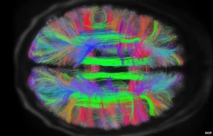
Tractography. This is a method of visualizing nerve fibers. It is performed using a magnetic resonance imaging (MRI) scanner. It shows how water molecules move along nerve fibers – white matter
tracts, and thus allows us to assess their integrity.
- Microscopy: Using powerful microscopes, scientists can examine tiny details, such as the work of neurons inside the brains of animals during experiments.
- Machine learning methods. Algorithms help process large volumes of data on neural connections and find patterns in them.
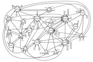
- 3D modeling. To better understand how the brain works, scientists create special models. In them, neurons are depicted as dots (nodes), and their connections are depicted as lines.
- Such models allow us to see how neural networks are structured and work, and also show how the brain structure changes over time.
Connectome research projects
Early works
Scientists took their first steps in studying brain connections in the second half of the 20th century. In 1986, a group led by British biologist and head of the Cambridge University Molecular Biology Laboratory Sydney Brenner compiled the first complete map of the nervous system of the small worm C. elegans. It has 302 neurons, so it was technically possible to map all the connections.
The C. elegans map showed the pathway a signal takes within the organism to trigger a particular behavior, such as a response to egg laying or touch.
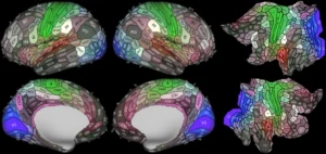
In 2008, a team of researchers led by Professor Patrick Hagmann, director of the Connectomics Laboratory at the University of Lausanne, visualized the connections between areas of the human cerebral cortex for the first time.
A map of connections between areas of the cerebral cortex (macroconnectome) obtained using MRI in humans (Photo: theplosblog.plos.org)
They noticed that the map of such connections differed in different people. This confirmed the hypothesis that each person’s brain is unique not only in the volume of gray and white matter, the thickness of the cortex, but also in the structure of connections between neurons.
The Human Connectome Project
Further conclusions required data from a large number of people. For this purpose, American scientists and the US Department of Health and Human Services launched the Human Connectome Project (HCP) in 2009 .
Researchers from different universities around the world collected data from thousands of volunteers using powerful MRI scanners. The result was an atlas that was unique in its detail and content. In it, scientists identified different areas of the brain based on their functions and connections with other areas. In each hemisphere, they counted 180 different segments, many of which were previously unknown.
Each of these areas can be thought of as a small department within a large company. For example, there is a department that analyzes visual information, next to it is one that is responsible for movement, another one is involved in planning, and so on. Different areas solve problems together.
As the HCP’s main phase concluded in 2021, new studies began to build on it. Some projects focused on studying the brains of depressed adolescents, while others focused on analyzing connectome changes in older adults. The research is ongoing, and its results may help develop new diagnostic and treatment methods for various diseases in the future.
Google Projects
In the last decade, Google has joined the study of neural connections. Together with leading universities and scientists, its specialists conduct large-scale research.
For example, in 2021, Google, in collaboration with Harvard University, completed a unique project called H01. Scientists processed more than 1.4 PB of data to create a highly detailed 3D map of a small fragment of the human brain. The researchers cut the organ tissue into thousands of ultra-thin sections and illuminated them with powerful electron microscopes. This made it possible to examine not only cells, but even individual contacts between them — synapses.

In 2024, Google and the Institute of Science and Technology Austria (ISTA) introduced a new way to study the connections between neurons using a light microscope. To see the smallest details, the scientists increased the size of the tissue using a special gel.
To do this, the specialists used brain fragments that were anonymously donated by patients during neurosurgical operations (for example, when treating epilepsy, a small section of the healthy cortex is sometimes removed to gain access to its deeper layers). Then the scientists soaked the brain tissue in hydrogel, which caused it to increase in size several times. This made it possible to examine individual connections between neurons using a regular light microscope. After that, using special dyes, they “illuminated” proteins, synapses, and other molecules in different colors.
This method helps to see how neurons connect to each other, as well as to distinguish between their types and functions. This allows for the rapid creation of detailed color maps of the brain, which immediately show both the connections and the features of the cells.
MICrONS Project
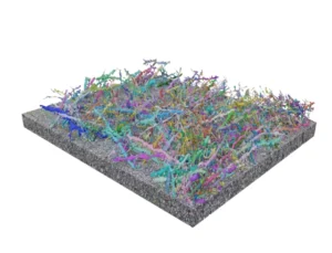
In April 2025, a team of researchers from the Allen Institute, Baylor College of Medicine, Princeton (USA) and other institutions presented the largest and most detailed map of the connections of the mammalian brain. It shows not only the structure, but also the work of neurons.
To map it, the scientists studied 1 mm3 of the mouse visual cortex and reconstructed more than 200,000 cells. Of these, about 82,000 were neurons. In this tiny area, they found about 500 million synapses.
At the level of individual types of neurons and their connections, the brains of mice and humans are similar . Therefore, scientists believe that analyzing the animal’s connectome will help to understand how failures in the functioning of neural circuits occur in Alzheimer’s disease or multiple sclerosis in humans. This may allow us to find new approaches to treatment.
EBRAINS infrastructure
The famous European brain mapping project Human Brain Project (HBP) started in 2013 and will be completed in 2023. Its goal was to develop new tools and technologies for studying the human brain.
As part of the project, the digital research platform EBRAINS (European Brain Research Infrastructures) was created in 2019. It includes databases on the structure and functions of the brain, modeling tools, and digital maps of the organ. EBRAINS allows neural connections to be modeled and helps specialists from different countries exchange scientific data
China’s Human Connectome Project
The Chinese Human Connectome Project (CHCP) is a scientific study launched in 2017. Within its framework, specialists analyze the connectome of East Asians, primarily the Chinese.
The main goal of the CHCP is to find out whether the brain is affected by environmental and lifestyle factors. Scientists have already presented some of the findings. Comparison of the CHCP data and the Human Connectome Project showed that the brains of people from different cultures – Chinese and Western – are generally similar. They have common features in structure, function, and connections. But there are also differences – in areas related to language and complex thought processes. These results will help to understand how culture and environment determine human behavior in the future.