


We have collected winning shots from international photography awards that show how photography helps scientists study complex natural phenomena
Nikon Small World
Nikon Small World is one of the most prestigious photomicrography competitions , established by Nikon in 1975. The uniqueness of the award is that the jury evaluates the works from both a scientific and an aesthetic point of view, thus creating a unique bridge between science and art.
Mouse brain tumor cells
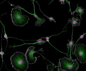
Differentiated mouse brain tumor cells (Photo: Bruno Cisterne with assistance from Eric Vitriol/Nikon Small World)
The photo shows mouse brain tumor cells, clearly showing the actin cytoskeleton (thin filaments that support the cell’s shape and facilitate its movement), microtubules (structures through which various substances move) and nuclei that store the cell’s genetic material. The author of the image is Dr. Bruno Cisterne in collaboration with Dr. Eric Vitriol of Augusta University, USA.
Scientists are studying how abnormalities in the cytoskeleton can lead to diseases such as Alzheimer’s and amyotrophic lateral sclerosis. “One of the key challenges in studying neurodegenerative diseases is that their causes are not fully understood,” explains Cisterne. “In order to develop effective treatments, we first need to understand the underlying mechanisms of these diseases. Our study plays an important role in finding this knowledge and opens up prospects for the development of new drugs.”
The image was taken at 100x magnification and was the winner of the Nikon Small World 2024 competition.
The optic nerve of a rodent
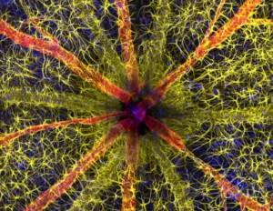
The winner of the 2023 Nikon Small World competition is an image of a rodent’s optic nerve taken by Hassanain Kambari and Jaden Dixon of the Lions Eye Institute in Australia. The image, taken at 63x magnification, shows a section of the rodent’s optic nerve head, the point where the optic nerve exits the eyeball.
Special fluorescent dyes are used to visualize collagen fibers (green), nerve cells (red) and the cell nucleus (blue). This image plays a role in the study and treatment of diabetic retinopathy, a retinal damage that occurs with diabetes.
“The visual system is a complex and highly specialized organ, and even relatively minor disturbances in retinal blood flow can lead to catastrophic vision loss. I decided to enter the competition to demonstrate the complexity of retinal microcirculation,” Kambari shares.
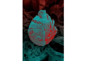
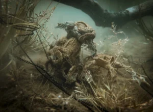
Gecko embryo foot
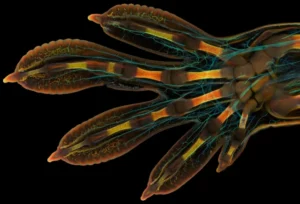
The photo shows the paw of a large Madagascar gecko embryo, its hand is only 3 mm long. The authors of the photo are scientists from the University of Geneva, Dr. Grigory Timin performed the work under the supervision of Dr. Michel Milinkovic. The image is artificially colored: the nerves are highlighted in blue, and the bones, tendons, ligaments, skin and blood cells are in warmer colors.
The shooting method used was confocal microscopy, which allows obtaining clear and high-contrast images by using point lighting and eliminating scattered light. In total, several hundred detailed images were taken, which were then combined into a single image. The entire process took about two days and required almost 200 GB of data.
The photo was the winner of the Nikon Small World 2022 competition.
Welcome Photography
The Wellcome Photography Prize is a photography award organised by the Wellcome Foundation, a British foundation dedicated to health research. The foundation was founded in 1936 with money left as a legacy by pharmaceutical magnate Henry Wellcome.
The competition includes several nominations, one of which is “Wonders of Scientific and Medical Visualization”.
Cholesterol in the liver
The 2025 winner in the Wonders of Scientific and Medical Imaging category is a photograph of cholesterol in the liver. The image shows cholesterol crystals (blue) inside a liver cell (purple). When cholesterol changes from a liquid to a crystalline state, it can build up in blood vessels and damage them, leading to heart attacks and strokes.
Scientific photographer Steve Gschmeissner created the image using an electron microscope, which allows one to see tiny structures with very high resolution.
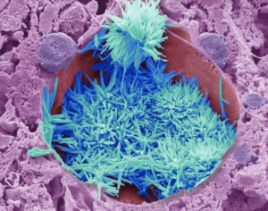
Triatomine bugs
Pictured is a triatomine bug, common in Latin America. The bite of this insect is dangerous to humans – it is a carrier of Chagas disease.
Chagas disease can lead to serious heart and digestive problems, especially if left untreated. It most often affects low-income people in rural areas of Latin America.
The photograph was taken using a cryoscanning electron microscope, and thanks to artificial coloring, individual cellular structures can be clearly seen.
Photo by Ingrid Augusto, Kildare Rocha de Miranda and Vania da Silva Vieira, researchers from Brazil who study the triatomine bug and hope their work will lead to a better understanding of how to combat Chagas disease.
Air Pollution
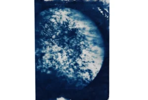
Polluted air on Brixton Road in London (Photo: Marina Vitaglione/Wellcome Photography Prize 2025)
The photograph shows fine particles, the most common air pollutant. Artist Marina Vitaglione, together with scientists from Imperial College London, collected air samples from different areas of the city. Vitaglione photographed these samples under a microscope and created prints using hand-made analogue printing with the addition of iron salts. This is why the colour of the photographs acquired a blue tint.
The photograph shows specimens collected from Brixton Road in south London.
“Pollution levels in central London have fallen over the past four years, but a quarter of roads still exceed legal limits for nitrogen dioxide (mostly from diesel engines) and millions of Londoners continue to breathe polluted air. This work aims to visualise the ‘invisible killer’,” says Vitaglione.
Royal Society Publishing Photography Competition
The Royal Society is a British scientific organization founded in 1660. Its mission is to advance science and disseminate scientific knowledge. One of the Royal Society’s modern initiatives is the annual scientific photography competition.
Shark attack
‘Hunting from Above’: A school of fish surrounded by sharks (Photo: Angela Albie. Drone pilot August Paula/Royal Society Publishing Photography Competition)
The shark photo is called “Hunting from Above.” It shows a large school of small fish confronting four young blacktip reef sharks. The image was taken by a drone off the coast of the Maldives.
This photo is part of a scientific study by biologist Angela Albi from the Max Planck Institute in Germany. Albi studies the interactions between blacktip reef sharks and schools of fish in the Maldives. “In this image, the shark on the left suddenly goes from swimming calmly in a school to starting to hunt. It stands out because of its posture,” Albi says . Biologists study photos and videos to understand how sharks hunt and how other fish react to them.
The photograph won first place in the Behaviour category and overall the 2024 Royal Society Publishing Photography Competition.
Constellation Cassiopeia
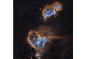
The Heart and Soul are two nebulae located in the constellation Cassiopeia, approximately 6,000 to 7,000 light years from Earth. The author of the photograph is Imran Sultan, a research fellow at Northwestern University in the United States. He spent almost 14 hours photographing these nebulae to capture their details and features. The photo won in the Astronomy category.
“My astrophotography has given me many opportunities to make astronomy more accessible to a wider audience. The fact that astronomers observed these two nebulae and saw a ‘heart’ and a ‘soul’ in them highlights the human element in astronomy,” Imran says .
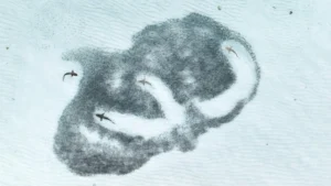
Common toads during the mating season
The photograph shows common toads during the breeding season, gathered in large numbers in shallow water.
“This photo was taken in the spring, when I was collecting eggs for experiments with my research team,” says the author of the photo, Ovidiu Dragan, a PhD student at Ovidius University in Constanta. “The whole area was literally overflowing with toads desperately trying to mate. What is especially interesting is that the second toad from the top is a male green toad, not a common toad. He was trying to mate with another species with which they coexist in their natural habitat. This behavior in the mountains was a big surprise for us.”
The photo won second place in the Behavior category. The photo was taken with a phone.
#ScientistAtWork
#ScientistAtWork is an annual international photo contest organized by Nature magazine. Its goal is to showcase scientists at work, whether in a lab, a park, the taiga, fjords, or even Antarctica. The organizers encourage researchers to share photos of their workdays and promise cash prizes to the winners.
Biologists in northern Norway
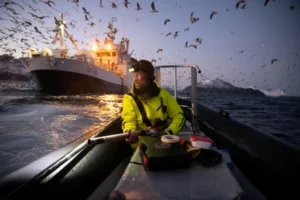
The winner of Nature’s Scientist at Work photo contest in 2025 is a photo by biologist Audun Rikardsen of his work in a fjord in northern Norway. A team of scientists from the University of Tromsø monitors the migration of herring, which attracts killer whales and humpback whales. They tag the whales with satellite tags, which are deployed with an air gun, to track their movements.
The tags collect data on the whales’ locations, as well as recording dive parameters, duration and depth. Scientists also often perform biopsies, taking tissue samples to monitor the whales’ health.
The work keeps researchers in close proximity to the animals. “You can actually feel their [the whales’] breathing,” says biologist Emma Vogel, who took the photo. “And you hear them before you see them, which is always amazing.”
Defenders of Frogs
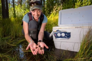
This photo is another ScientistAtWork winner. It shows ecologist Kate Belleville of the California Department of Fish and Wildlife holding baby frogs.
In the Lassen National Forest in northern California, a team of scientists and volunteers capture young frogs to bathe them in an antifungal solution. The solution kills the chytrid fungus that is causing mass die-offs of amphibians around the world. After the treatment, the frogs are released back into the wild.
The froglets are also implanted with elastomer tags with a unique combination of colors. These tags form a code that glows under ultraviolet light.
As the researchers note, it is extremely difficult to notice the small frogs – if you do not know what you are looking for, they can easily be mistaken for scurrying crickets. Therefore, the work requires special caution and attention.